
String Art: Difference between revisions
(Created page with "=String Art: Axon Tracts in the Spinal Cord= These string boards are based on classic 1970's string art. Students use their hands and coloured wool to trace and learn several ...") |
No edit summary |
||
| (5 intermediate revisions by the same user not shown) | |||
| Line 7: | Line 7: | ||
* Spinocerebellar tract (sensory) | * Spinocerebellar tract (sensory) | ||
<gallery mode="nolines" widths=200px heights=200px> | |||
StringArt01.jpg|1 | |||
StringArt02.jpg|2 | |||
StringArt03.jpg|3 | |||
StringArt04.jpg|4 | |||
StringArt05.jpg|5 | |||
</gallery> | |||
== Downloadable files == | == Downloadable files == | ||
Composite image file: StringArtImage.pdf | Composite image file: [https://anatomy-teaching-models.wiki.otago.ac.nz/images/f/f8/StringArtImage.pdf String Art Image] | ||
Cheat sheet: String_Art_Spinal_Neuron_Tracts. | |||
Cheat sheet: [https://anatomy-teaching-models.wiki.otago.ac.nz/images/1/11/String_Art_Spinal_Neuron_Tracts.pdf String Art Spinal Neuron Tracts] | |||
== Production Information == | == Production Information == | ||
| Line 21: | Line 29: | ||
This work is licensed under a [http://creativecommons.org/licenses/by-nc-sa/4.0/ Creative Commons Attribution-NonCommercial-ShareAlike 4.0 International License] | This work is licensed under a [http://creativecommons.org/licenses/by-nc-sa/4.0/ Creative Commons Attribution-NonCommercial-ShareAlike 4.0 International License] | ||
Latest revision as of 04:47, 10 August 2016
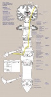
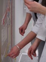
String Art: Axon Tracts in the Spinal Cord
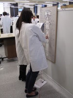
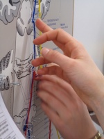
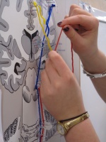
These string boards are based on classic 1970's string art. Students use their hands and coloured wool to trace and learn several axon tracts:
- Spinal reflex arc
- Corticospinal tract (motor)
- Dorsal column tract (sensory)
- Spinothalamic tract (sensory)
- Spinocerebellar tract (sensory)
1
2
3
4
5


Downloadable files
Composite image file: String Art Image
Cheat sheet: String Art Spinal Neuron Tracts
Production Information
Stylised images of different levels of the brain and spinal cord (based on actual sections) were drawn out and the various tracts and nuclei marked out within them. The final composite image was printed out on adhesive plastic and stuck onto board. Nails were hammered in - large headed nails for cell bodies and finer nails for waypoints. A cheat sheet was also produced to assist the students.
Creators: Dr Marilyn Duxson, Dr Ping Liu and Dr Brad Hurren (academic support), Fieke Neuman and Robbie McPhee (initial drawings and final artwork), Alex Witherow (wooden boards). All from Department of Anatomy, University of Otago.
Keywords: Teaching, Anatomy, Neuroanatomy, Axon, Neuron, Brain, Spinal Cord, Reflex, Motor Control, Sensation, Science, Biology
![]()
This work is licensed under a Creative Commons Attribution-NonCommercial-ShareAlike 4.0 International License