
GutTube: Difference between revisions
mNo edit summary |
No edit summary |
||
| Line 4: | Line 4: | ||
Long rectangles (80mm x700mm) were cut from artificial fur fabric. The two long edges were sewn together and overlocked, with the fur on the inside. The ends were left open so that marbles could be introduced. Plastic cling wrap was used to simulate the gut mesentry, with coloured yarn to simulate arteries, veins, lymphatics and nerves within the mesentry. | Long rectangles (80mm x700mm) were cut from artificial fur fabric. The two long edges were sewn together and overlocked, with the fur on the inside. The ends were left open so that marbles could be introduced. Plastic cling wrap was used to simulate the gut mesentry, with coloured yarn to simulate arteries, veins, lymphatics and nerves within the mesentry. | ||
<gallery mode="nolines" widths=200px heights=200px> | |||
<gallery mode="nolines" widths= | |||
Guttube01.jpg|1 | Guttube01.jpg|1 | ||
Guttube02.jpg|2 | Guttube02.jpg|2 | ||
| Line 27: | Line 18: | ||
Guttube12.jpg|12 | Guttube12.jpg|12 | ||
</gallery> | </gallery> | ||
'''Creators:''' Dr Ruth Napper, Dr Rebecca Bird and Fieke Neuman. All from [http://www.otago.ac.nz/anatomy Department of Anatomy], [http://www.otago.ac.nz University of Otago]. | |||
'''Keywords:''' Teaching, Anatomy, Gut, Peristalsis, Mesentry, Science, Biology | |||
[[Image:CC.png|link=http://creativecommons.org/licenses/by-nc-sa/4.0/]] | |||
This work is licensed under a [http://creativecommons.org/licenses/by-nc-sa/4.0/ Creative Commons Attribution-NonCommercial-ShareAlike 4.0 International License] | |||
Latest revision as of 04:46, 10 August 2016
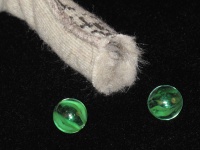
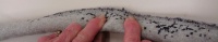
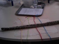
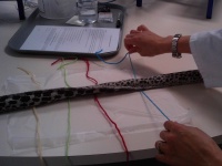
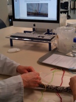
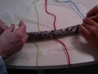
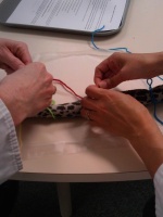
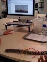
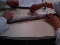
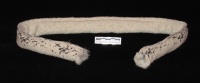
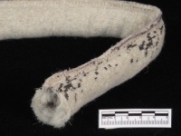
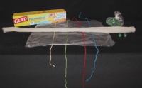
A simple rectangle of fun fur becomes a gut tube for first year health science students to understand how peristalsis can move food (marbles) from one end to the other. Plastic film and coloured wool simulate the gut mesentery containing blood vessels and nerves.
Production Information
Long rectangles (80mm x700mm) were cut from artificial fur fabric. The two long edges were sewn together and overlocked, with the fur on the inside. The ends were left open so that marbles could be introduced. Plastic cling wrap was used to simulate the gut mesentry, with coloured yarn to simulate arteries, veins, lymphatics and nerves within the mesentry.
1
2
3
4
5
6
7
8
9
10
11
12


Creators: Dr Ruth Napper, Dr Rebecca Bird and Fieke Neuman. All from Department of Anatomy, University of Otago.
Keywords: Teaching, Anatomy, Gut, Peristalsis, Mesentry, Science, Biology
![]()
This work is licensed under a Creative Commons Attribution-NonCommercial-ShareAlike 4.0 International License