
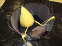
Female Pelvic Peritoneum (in a bucket)
This schematic model was developed to help third year medical students understand the relationships between the parts of the peritoneum of the female pelvis. The model shows organs, ligaments and peritoneum within the lesser or true pelvis i.e. only from the pelvic inlet down.
Click to enlarge images:


Production Information
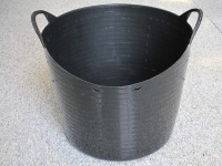
This model is built to fit inside a 26 litre flexible bucket (also known as flexitub). The anterior edge of the bucket is cut lower to give an idea of the anterior pelvic brim and a hot tool used to create openings for the anus, vagina, urethra and external inguinal canals. The hot tool is also used to mark out labia, clitoris and the perineal triangles.


The fabric parts of the model are made from tulle (aka fine bridal netting), coloured fabrics (pink for uterus, yellow for bladder and ureters, brown for rectum, white for ovaries, red for arteries) and string for the ovarian and round ligaments. The organs are stuffed with wadding and the whole assembly held in place with nylon fishing line.
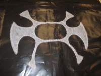
The pattern pieces (background grid is 100mm x 100mm):
Click to enlarge images
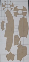
Peritoneum pattern pieces
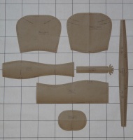
Uterus patten pieces
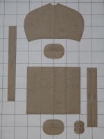
Bladder, rectum, ovaries, arteries pattern pieces


Completed organs:
Click to enlarge images
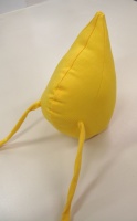
Bladder
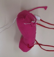
Uterus
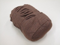
Rectum


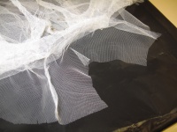
As the construction of the tulle peritoneum is complicated some of the important steps are illustrated below:
Click to enlarge images
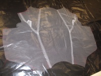
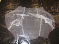
1. Join the two parts of the broad ligament to each other and then to the uterine peritoneum.
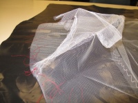
2. Stitch through the anterior broad ligament to create the mesosalpinx. Note that an opening is left for the fimbriae of the uterine tubes.
3. Wider view of step 2.
4. Preparing to sew on the two parts of the peritoneal base.
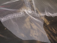
5. Note the wrinkle of fabric that will become the ureteric fold.
6. Join the peritoneal base to the uterine peritoneum/broad ligament.
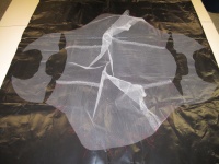
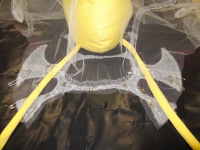
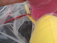
7. The position of the parietal peritoneum within the bucket. Sew the bladder peritoneum between the ends of the main parietal peritoneum. Note red thread marks for where the broad ligament will attach to the parietal peritoneum.
8. The 4 layers of the peritoneal base ligaments are sewn together.
9. Anterior and posterior parts of the peritoneal base are sewn separately to the base ligaments. Note that there is a gap of about 1cm between the anterior and posterior parts of the broad ligament as they rise up from the peritoneal base. Sew down the ureteric folds.
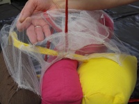
10. Sew the long curved edge of the pararectal fossa peritoneum to the posterior part of the peritoneal base.
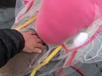
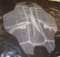
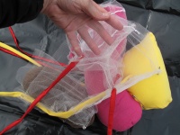
11. Gather and sew the main parietal peritoneum to the remaining edges of the pararectal fossa to create a pocket. The organs can be positioned inside their relevant cavities. Note hand in rectouterine pouch (pouch of Douglas)..
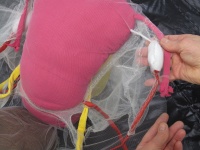
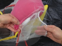
12. Sew the numbered marks on the edge of the broad ligament to corresponding marks on the parietal peritoneum. Make small holes in the tulle for the exits of the round ligament, uterine artery and ovarian artery.
13. Note how the ureter passes under the uterine artery.
14. Sew additional lines of stitching to delineate the mesovarium and mesosalpinx from the mesometrium.
15. The fold of peritoneum that covers the round ligament is exaggerated in this model compared to life.
16. Sew the organs together and sew the completed peritoneum to the organs.
17. Melt pairs of small holes through the bucket at appropriate points so that fishing line can be used to tie the peritoneum in place within the bucket.


Creators: Dr Latika Samalia (initial idea and academic support), Fieke Neuman (patterns, markings and sewing). Both from Department of Anatomy, University of Otago.
Keywords: Teaching, Anatomy, Medicine, Pelvis, Reproduction, Science, Biology
![]()
This work is licensed under a Creative Commons Attribution-NonCommercial-ShareAlike 4.0 International License